TLDR
- Skin is not just a bag around your bones. It plays three critical functions: protection, grip surface and load path, and a dense sensor network.
- Human skin repairs itself via a four-stage process (hemostasis, inflammation, proliferation, remodeling). Robotics is exploring self-healing polymers to replicate this, though modular design (wholesale swapping of damaged skin modules) may prove to be a more practical complementary approach for larger damage in most applications.
- Human skin has stiff friction ridges (the bumps in our fingertips) that bite into rough surfaces for traction, or soften via moisture plasticization to a rubber-like material that conforms to smooth surfaces.
- Humans have thousands of sensors per hand: four types of mechanoreceptors and two types of thermoreceptors that sense vibration, surface pressure, stretch, and temperature, enabling dexterous manipulation.
- Roboticists are exploring many sensing technologies (FSR, piezoelectric, optical GelSight Mini, DIGIT, magnetic AnySkin, microbarometers), but no single one fills the gap. Achieving parity requires integrating 2 to 3 sensor types and designing for serviceability.
Context
Robotic hands are currently a major focus in the industry. The industry has realized that dexterous hands will be critical to make robots general-purpose enough to interact with a world designed for humans. Current efforts largely focus on downsizing actuators to match human grip strength while remaining compact enough to fit in the hand and forearm. I applaud those endeavors and find them incredibly interesting, but there is an under-representation of focus on the challenges of making a robotic version of human skin.
There are levels of complexity here that rival the mechanical challenges of the robotic hand itself. Akin to the challenge of packaging actuators is the challenge of packaging the dense array of tactile and temperature sensors into a mechanically sound, easy-to-service grip surface. The role these sensors play in humans enables us to have spatial understanding of our world and to manipulate delicate tools and heavy objects. Some of our recent paper analyses, like Touchy Subject, the review of the VISK framework, have shown that multimodal learning, by adding tactile sensation, has significantly improved the robot's spatial awareness and object manipulation capabilities, which are current challenges for all of today's robotic policies.
Studying the human body provides critical insights for creating artificial systems. While this article focuses primarily on the skin around our hands, it also highlights broader anatomical principles that inform robotic design.
Skin as a Protective Outer Layer
The skin has three main layers: the epidermis, dermis, and subcutaneous layer (sometimes called the hypodermis). These layers remain separate but interlock through a high level of surface texture (not visible here but evident in later images).
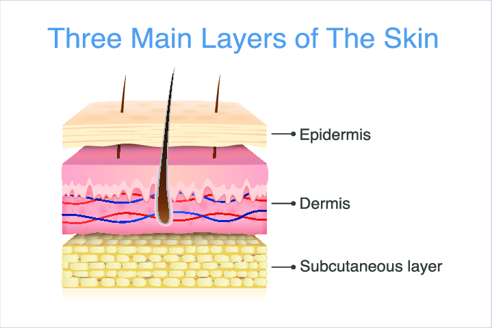
Both the epidermis and dermis play critical roles in the protective outer-layer functionality, which comprises the following attributes:
- Moderate surface toughness
- Water resistance
- Moderate tensile strength
- Self-repairing capabilities
Moderate Surface Toughness: A Primary Role of the Epidermis
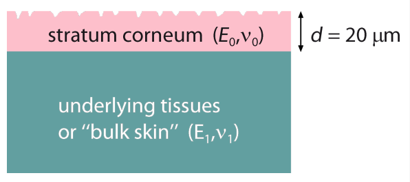
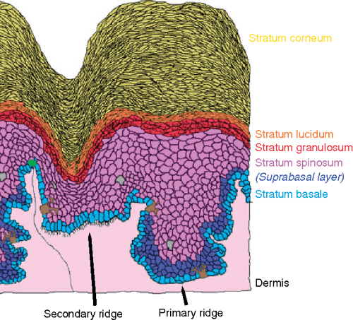
The epidermis is the skin's tough yet flexible outer shield. While it is paper-thin over most of your body, typically ranging from 0.03 to 0.1 millimeters, it undergoes a massive architectural upgrade on your palms and soles. In these high-friction areas, studies show that skin adapts to consistent stretching, rubbing, and friction by thickening to enhance abrasion protection and grip. In these high-impact areas, the total epidermal thickness can reach 1.5 mm. This is largely because the stratum corneum, the outermost layer of toughened cells, expands from a mere 20 micrometers elsewhere to as much as 700 micrometers (0.7 mm) on your hand to withstand the mechanical stress of heavy work cycles.
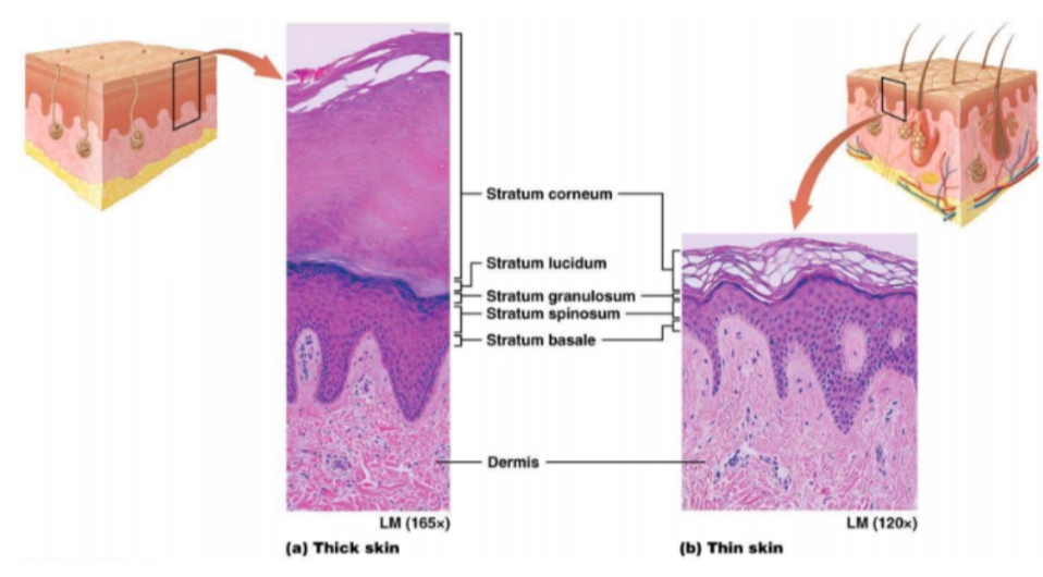
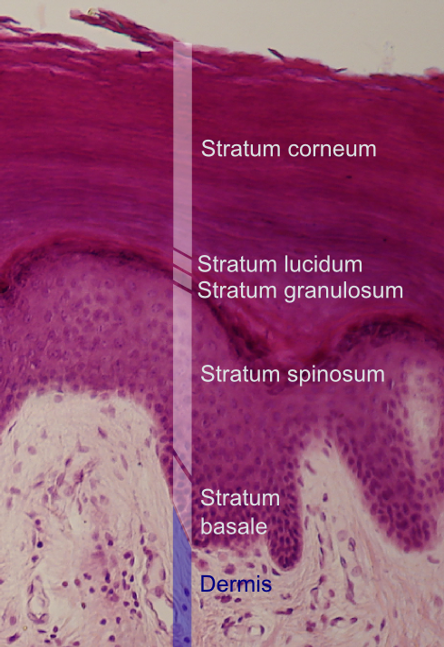
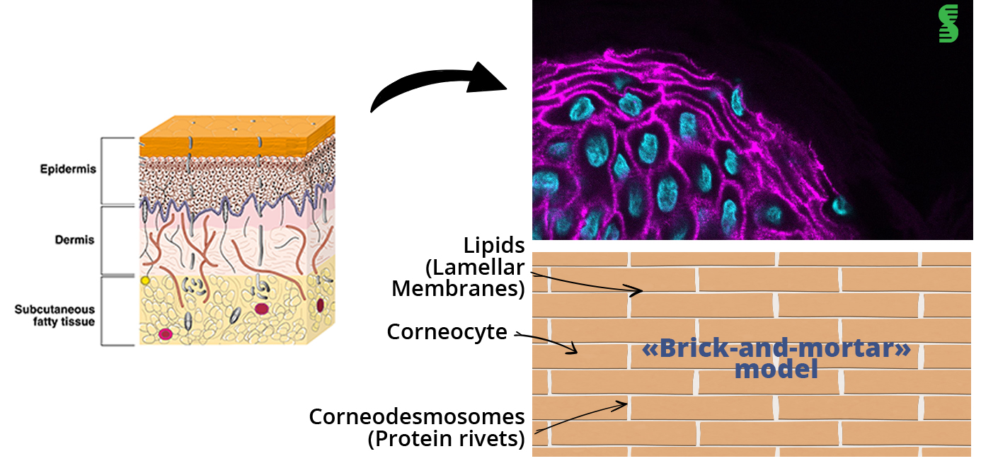
The epidermis is made up of keratinocytes, cells that produce keratin, the tough, fibrous protein in hair and skin that makes it strong and resilient. The stratum basale, at the base of the epidermis, generates these cells, which feature small fingers that interlock more tightly as they move towards the surface. Subsequent layers generated below push each layer upward. You can see the mess of cell fingers as the lighter pink area in the stratum spinosum layer; the dark areas are the cell bodies. By the time they reach the stratum corneum, the cells are self-sacrificial: dead, flattened, and devoid of a nucleus, internally reinforced with keratin fiber bundles, and their fingers tightly interlocked with those of neighboring cells via desmosomes, a sticky protein. As the outer layers are damaged or age, they dry out and flake off, making space for new layers below. Cells in the epidermis live about 30 days from generation to floating in the breeze as skin flakes.
Water Resistance
In addition to the keratinocytes producing keratin internally, they also release a lipid byproduct externally. This lipid release explains why our skin feels oily. These lipids pack tightly between the cells and the interwoven fingers, acting as mortar between bricks or pine tar between planks in the hull of wooden ships to create a hydrophobic, watertight barrier.
Moderate Tensile Strength: Courtesy of the Dermis
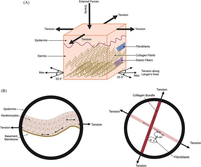
The dermis is the infrastructure layer of the skin; it is plumbed with blood vessels, nerve endings, and eccrine glands (sweat glands). It acts as the active core layer, composed of fibrous connective tissue, with bundles of collagen and elastin fibers. These fibers naturally form a wavy structure. Figure 3 shows these waves as faint pink bundles in the dermis. This allows high flexibility under normal movement and strain, but as the skin experiences higher tensile loads in any direction, the waves tighten and bear most of the load, preventing the skin from tearing apart.
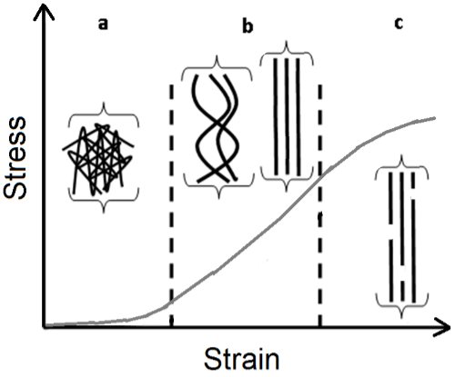
The dermis provides the structural component of the skin. In 2012, researchers measured the skin's ultimate tensile strength (UTS) at 27 (+/- 9.3) MPa in back skin samples [1]. This is nearly twice that of rubber (16 MPa) but only 75% of the strength of ABS (40 MPa) [2]. Our palms are likely stronger than this, though I could not find specific research on the tensile strength of the palm dermis. It's the interlocked waves of the collagen bundles in the dermis that bear the majority of this load, like the glass fibers inside of glass-filled injection-molded parts. While the epidermis provides material toughness and cut resistance, the dermis provides the material's high tensile strength and rupture resistance. This is why a surgeon will anchor sutures into the dermis when closing up a wound, since the epidermis alone would not be able to bear the load and could tear.
Active Self-Repair Capabilities
The second, and arguably most important, feature of the epidermis is its ability to repair itself. It constantly creates new cells to replace older or damaged ones, giving us a self-regenerating surface so that minor nicks and scratches aren't debilitating for long. This is perhaps one of the most challenging features of human skin to replicate in robotics.
When a wound breaches the protective epidermis and penetrates the vascularized dermis, the body initiates a complex four-stage repair process. The immediate priority is hemostasis, which plugs the leak and prevents a catastrophic drop in fluid pressure. Platelets from the bloodstream clump at the rupture and trigger a clotting cascade that polymerizes fibrin, creating a temporary biological glue to stabilize the site. With the leak sealed, the body enters the inflammation phase. Local blood vessels dilate to allow white blood cells, such as neutrophils and macrophages, to flood the area and act as autonomous biological scrubbers, digesting foreign debris and bacteria. Once these cells sanitize the site, the proliferation phase begins. Fibroblasts migrate into the temporary fibrin scaffold and synthesize a temporary bundle of collagen fibers to bridge the gap, while new blood vessels sprout to supply the massive energy demand of reconstruction. Finally, during the remodeling phase, more methodically arranged collagen bundles slowly replace the hastily arranged collagen. The body aligns these new fibers along lines of mechanical stress to optimize the load path, a process that can take weeks to years, depending on the severity of the injury.
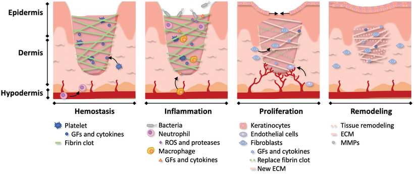
The depth of the damage entirely dictates the final material makeup of the repaired skin. A shallow surface scrape that only damages the epidermis will bypass the hemostasis and inflammation phases because there are no blood vessels to sever or clear. Instead, the local keratinocytes in the epidermis simply detach from their basal layer, migrate across the shallow defect, and proliferate to fill the void, resulting in perfect regeneration of the original tissue with no loss of mechanical properties. However, a deep cut that severs the dermis's complex honeycomb matrix forces the body to patch the structure rather than regenerate it. The resulting patch is a scar. In scar tissue, fibroblasts lay down dense collagen fibers in parallel bundles rather than the original multiplanar weave. This makes the tissue strong in one specific direction but significantly weaker in others. Furthermore, the body does not synthesize new elastin fibers or rebuild complex appendages like sweat glands during this patch job, leaving the scarred area permanently lacking the flexibility and moisture-regulating grip surface of the original skin.
Robotic Equivalents for Protective Skin
Currently, many robotic skins focus purely on aesthetics, like Xiaomi's famous presentation showing actuators beneath a silicone shell, or they rely entirely on hard plastics. While a plastic shell is probably sufficient for most of a robot's body, the unique grip and sensory requirements of the hand mean a rigid shell will not cut it.
Robot skin has slightly different baseline requirements than human skin. Robots are resilient to biological infections, rely on electrical circuits rather than microfluidic networks, and are built from actuators and components that often carry their own ingress protection ratings. Because of this, we do not necessarily need robotic skin to serve as the primary barrier against external elements. However, as we add high-density tactile and sensory modalities to electronic skins, the miniaturization of these components will leave no space for individual environmental protection. In that scenario, the skin's overall water resistance will become a necessary attribute to achieve the sensor density seen in the human body.
Fortunately, robotics can draw from a vast body of polymer material science. The search for higher-performance tires and high-temperature seals has led to incredibly strong TPUs that perform well in both rubberized and hard-plastic forms. Engineers can also utilize a large variety of foams and rubbers as flexible materials, perforating them with gaps for wires while maintaining a grippy, water-resistant surface.
For material repair, the most obvious solution for humanoids is to emulate human operation. Researchers are heavily investigating self-healing materials. One promising example comes from Bao Lab at Stanford University, which created a silicone polymer with reversible hydrogen bonds that allow breaks to reconnect at room temperature in a matter of minutes. Terasaki Institute shows promising use of silver nanowires and dynamic disulfide bonds that heal across a wide array of temperatures and humidities. Aalto University has shown that clay/polymer gel composites can have high surface stiffness, be flexible, and self-repair within 24 hrs of injury.
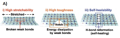
However, unlike humans, robots have other options for repair, including patch-and-repair systems or outright part replacement. A group at the University of Tokyo has found a way to grow collagen-based skin over the robot's surface and to repair surface tears using a collagen sheet patch and 7 days of incubation. While the 7-day incubation period required to fully cure the patch makes this commercially unviable, it does suggest an interesting idea: what if we could use external intervention to repair our robot's skin? It could be done by a human, another robot, or a dedicated tool, but there is no inherent need for the robot to repair itself, unless robots are deployed in a very remote location, such as the moon, where access to these tools is impossible.

While these self-healing polymers are incredible feats of materials science, the material costs, limited supply chain, and time required to achieve sufficient self-healing may prove impractical and unnecessary in most, if not all, cases. For heavy-duty applications, the most viable path will likely bypass biological mimicry in favor of modular design. Much like replacing a scratched trim piece in your car's interior, maintaining a robotic hand will involve unbolting the compromised sensory layer and swapping in a fresh, fully calibrated module to get the system back online immediately.
We will have to rely on replaceable skin modules designed for rapid serviceability. If we do not design for service, humanoid robots will remain financially unobtainable, much like not being able to afford a car because you have to replace the entire vehicle every time it needs an oil change. This is how we handle part failures in most machines and indeed most robots. I think this is the most likely way we will handle this challenge in general purpose robots, including humanoids. Expect heavy iteration on volar skin replacements in the near future.
Skin as a Grip Surface & Load Path
Treads of the Hands
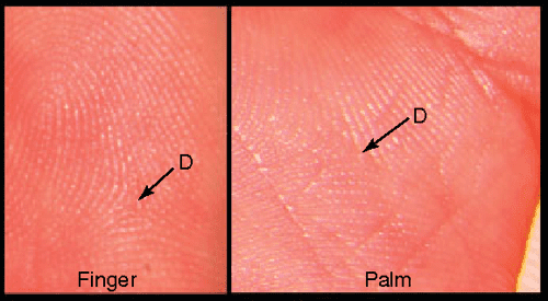
The skin on the hand is not as smooth as a baby's bottom; pores and ridges cover its surface. These ridges allow the skin to interlock with the irregularities of rough surfaces, very much like the tread of a tire.
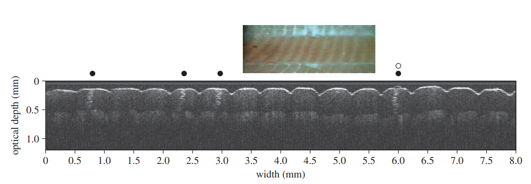
The most well-known examples are the papillary ridges on the palm and fingertips, which form our unique fingerprints. They serve dual purposes: biting into the objects we grab to provide traction and acting as antennas to transmit delicate contact and pressure to the haptic sensors deep below.
The outermost portion of the stratum corneum acts as a rigid shield with a Young's Modulus between 1 and 3 GPa when dry. This stiffness makes the surface behave like hard plastic, providing puncture and abrasion resistance during heavy work. The layers directly beneath this shell remain in a much softer state due to their higher moisture content, creating a composite structure that distributes pressure and protects sensitive internal tissues from point loads.
The hand can switch its material state from a hard protective layer to a high-traction surface through a process called moisture plasticization. Hundreds of eccrine (sweat) glands embedded along the friction ridges drive this transformation, acting as biological irrigation ducts. These glands deliver small amounts of sweat that infiltrate the keratin, reducing the elastic modulus by as much as 1,000 times. As the tissue softens, it transitions to a compliant, rubbery state, allowing the fingerprint ridges to flatten and conform to the microtexture of an object. This shift can increase the real contact area from 25% up to 50%. [3]
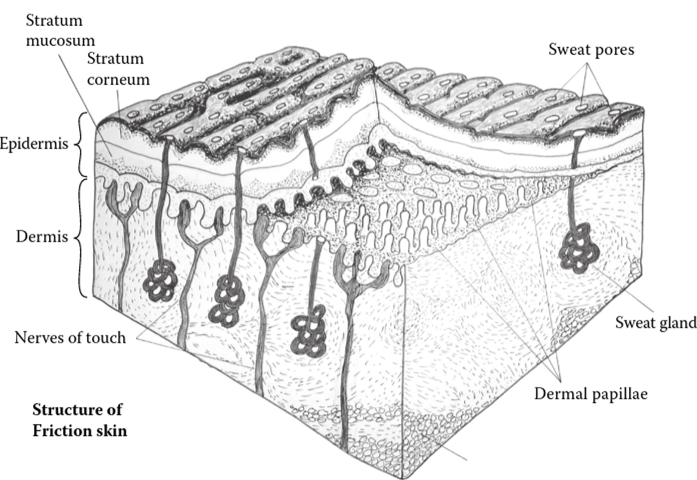
A passive microfluidic feedback loop regulates this system, preventing the fingertip from becoming oversaturated and slippery. As the ridges become soft and compliant under grasp, they flatten, physically sealing the sweat pores on their crests. This halts moisture release once the ideal hydration level is reached. Additionally, the valleys between the ridges act like tire treads, draining excess liquid. This mechanism resembles a high-performance tire: when cold and rough, the ridges dig in to protect against tears; as they heat up (or get wet), they soften to increase surface adhesion exactly where needed.
Structural Anchoring for Torque Resistance
The skin is not simply a bag around the human body's muscles and bones. It is a critical part of the load path between our bodies and the world. Together, we will explore the load path from inter-layer shear resistance to fascial structures and, finally, to the skeletal structure.
The dermis anchors the first layer, the epidermis. The border between the layers is full of interlocking ridges that accomplish this mechanically. On the epidermis, the friction ridges extend down towards the dermis in a major and minor set of ridges, interlocking with the dermal papillae. The primary set is deeper, running directly below the friction ridge, and contains the duct for the sweat gland. The secondary ridge lies beneath the channel and is shallower than the primary ridge. The receiving side is the dermal papillae, a 3-dimensional set of arms and hooks in the dermis that attach to the epidermis, much like Velcro hooks attach to loops on the epidermis.
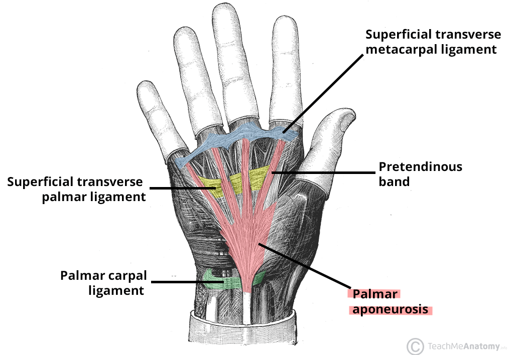
The secondary step happens under the palm, where the dermis anchors into a thick, triangular layer of fascia called the palmar aponeurosis. This structure ties into the ligaments woven around the base of each of the four fingers, depicted in blue, and connects to the ligament at the base of the wrist, not shown below. It consists primarily of collagen fibers that organize into a network of longitudinal, transverse, and vertical bundles to provide substantial tensile strength to the hand structure.
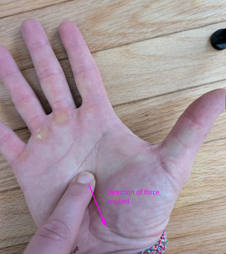
If you use your left index finger to rub the palm of your right hand, you will notice the skin barely moves, as if it's fixed. This stability results from anchoring to the palmar aponeurosis. Now, if you take that finger again and pull it towards your wrist, make sure it doesn't slip. You will feel tension at the base of your fingers, both on the palm side and on the back of the hand. You are starting to feel the load path from the skin of your palm into your skeletal structure, since the palmar aponeurosis ties into ligaments that loop around each of the four fingers. This fascial layer attaches to the dermis, with a honeycomb of fibers that create multiple load paths for tension to travel from your grip to your skeletal structure.
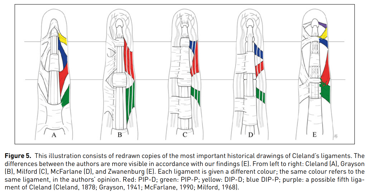
The fingers have similar structures called Cleland's ligaments, which criss-cross to tie the finger skin down to the joints in both the proximal (palm) and distal (fingertip) directions. The colors below highlight the different bundles; both sides of the finger mirror each bundle. This provides the load path from the skin of the finger pad down into the skeletal structure. It is also worth noting that the Grayson ligament, depicted as an uncolored horizontal wrapping in C and D, connects the fatty pads in the fingertips and bases of the fingers, which bear much of the load when handling heavy objects.
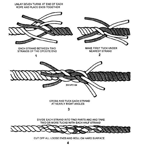
Connections between the fascial layers occur via fascial fiber bundles (septa) that interweave with the collagen matrix, forming a honeycomb-like pattern in the dermis. This structure physically resembles the short splice of two ropes, interweaving their strands for maximum strength.
Shock Absorbers and Load Distribution
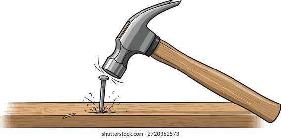
The body handles many high-impact scenarios, from everyday activities like running, jumping, or catching a ball to specialized activities like shooting a rifle or swinging a hammer. All of these are high impact, north of 1g, and could stress and tear muscle, tendon, and fascia alike. However, the energy-absorbing and load distribution capabilities of the skin and underlying muscle tendons render them harmless.
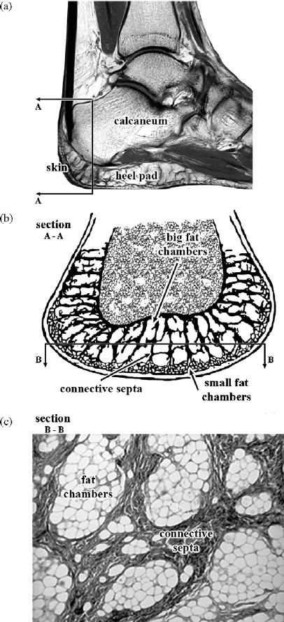
Beneath the fingers and palms live specialized fibrous fat pads called volar fat pads. When you grasp a hard object, the fat cannot simply squish out of the way; instead, the fibrous walls (septa) trap the fat globules, acting as hydraulic shock absorbers. These fat pads compress, turning 35%-60% of the kinetic energy into heat while safely transmitting the remaining forces into the skeleton, preventing nerves and blood vessels from being crushed.
Robotic Equivalents for Grip Surfaces and Structures
We can draw on extensive material science for robotic equivalents. Composites blending high-grip rubbers, structural components, and flexible adhesives could replace ligaments to secure a grippy surface to a metallic chassis.
If woven, tendon-based actuation takes off, it may naturally lead to woven anchor points for grip surfaces. Since human ligaments move the grip surface in tandem with hand flexion, a woven anchor point might be the only way to replicate this without adding unnecessary degrees of freedom.
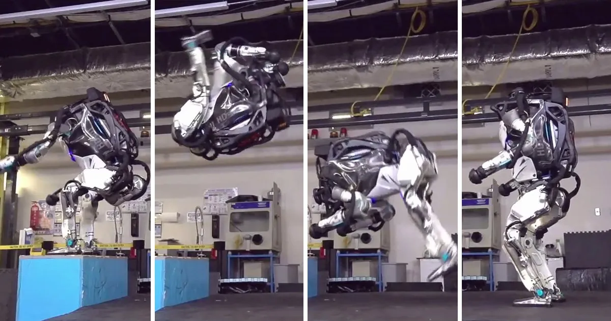
For shock absorption, TPU has become quite popular. Boston Dynamics famously uses TPU in the knee joints to help soften the blow. And I would be surprised if they don't start adding it to the base of the heel and the palm of the robotic hand, where it seems to be very impactful, pun intended, for human shock absorption.
Multimodal Sensor Network
The skin on our hands contains thousands of mechanoreceptors (haptic), thermoreceptors (temperature), nociceptors (pain), and more. By volume, the mechanical, temperature, and pain sensors outweigh the rest, so that is our focus.
Mechanical Sensation
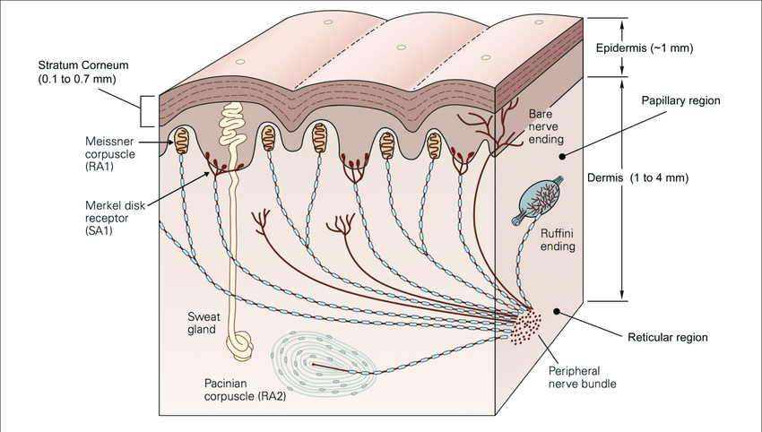
Haptic sensation requires a highly specialized, multi-layered approach. We have four different types of mechanoreceptors that react to different events and frequencies, giving us an incredibly wide dynamic range.
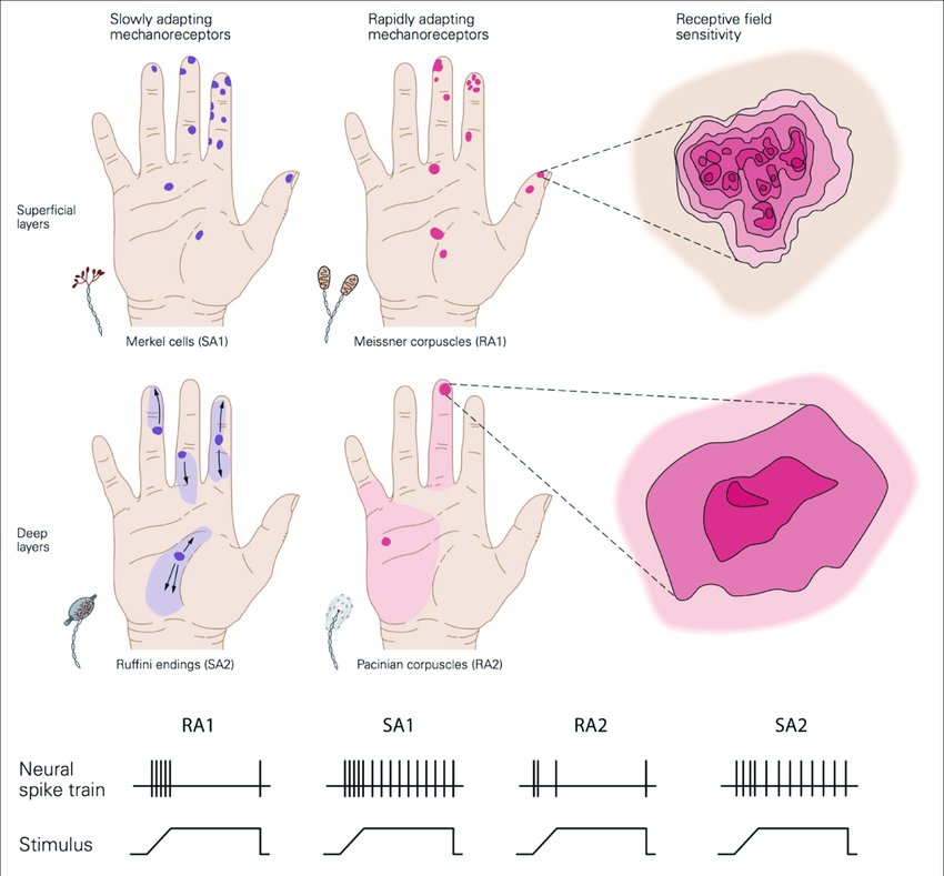
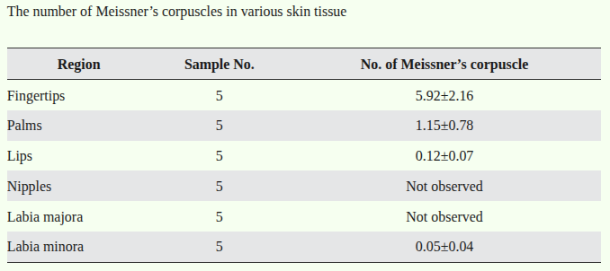
- Meissner Corpuscles (10-50 Hz): Located at the top of the dermis, near the dermal papillae, clustered around the areas with friction ridges. These detect the start of contact. They light up initially, then normalize and fade into the background. The fingertips contain roughly 6 per mm², while the palms contain 1 per mm². [4]
- Merkel Disks (Static Pressure): Clustered at the base of the fingerprints, these are slow-adapting sensors with sharp receptive fields. They help us determine forms, shapes, and spatial details, operating at forces from 3 mN to roughly 5 N.
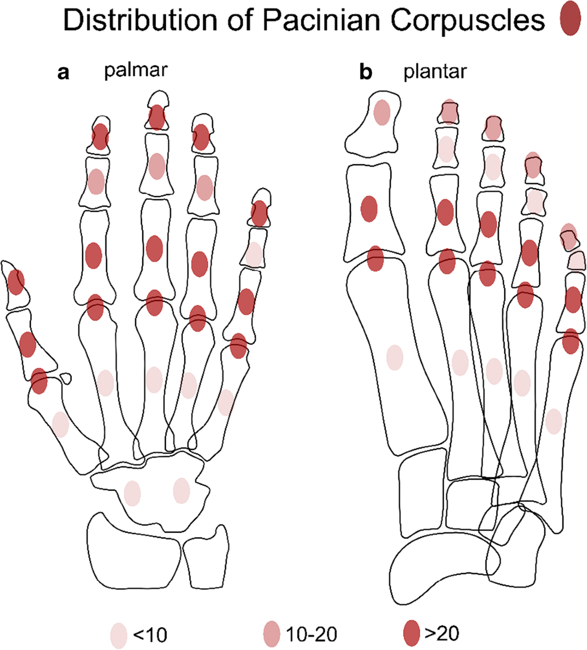
- Pacinian Corpuscles (60-320 Hz): Buried deep in the hypodermis, these detect high-frequency vibrations. Layered like an onion, corpuscles filter out low frequency with each subsequent layer, allowing us to detect a one-micron scratch or sense what a tool is touching at its tip. This is known as haptic hyperacuity. Pacinian corpuscles are the most sparse mechanoreceptor; the average person has about 150-200 across the entire palm and an additional 50-60 per finger. [5]
- Ruffini Endings (Stretch/Shear): Positioned to detect lateral tension and skin stretch, these are directionally sensitive and provide continuous feedback, which is critical for using tools like a wrench or writing with a pen.
The array and multi-modality of these sensors unlock incredible spatial awareness:
- Force Detection: Meissner Corpuscles detect the onset of contact, allowing our brain to perceive the material. The Merkel disk measures constant pressure to determine the applied force, which saturates at around 5N per fingertip once the fibrous fatty pad fully compresses. Beyond that, we rely on our muscular strain to gauge heavy loads.
- Shape Detection: Based on how the array of force detection sensors light up, we can detect surface features such as bumps and lines.
- Material Identification: The Meissner Corpuscle for rough textures, the Pacinian Corpuscle for fine textures, and the thermoreceptors, discussed below, combine to enable material identification. An alternative pathway to the brain combines these sensors with our visual expectations, giving us an innate ability to instantly identify the material we are touching.
- Skin Tension / Hand Torque: Then come the Ruffini endings, slowly adaptive and directionally sensitive, measuring the direction of skin tension. They provide continuous feedback as the load is applied, which is critical for using tools like a wrench or a screwdriver, or the feeling of writing with a pen.
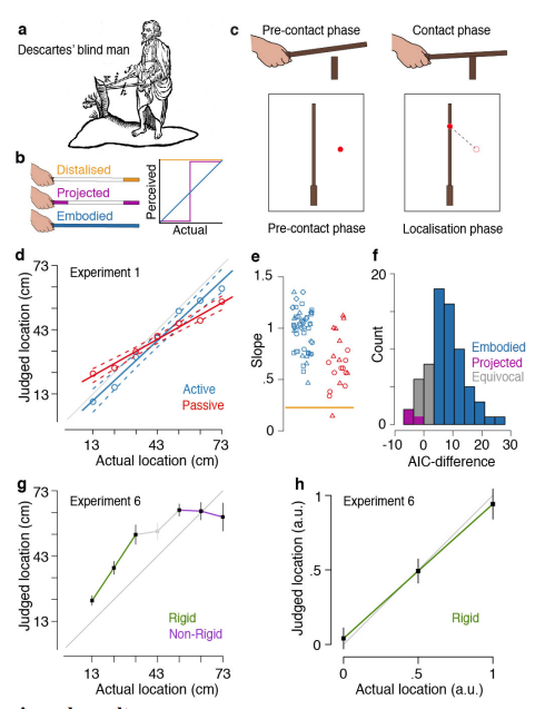
- Mode Estimation / Tool Proprioception: The Pacinian corpuscle allows us to detect the vibrational modes of the tools we're using, based on our understanding of the material, shape, and how they vibrate in our hand. Studies show humans are significantly more accurate at estimating distance when we combine this sense with our eyesight, compared to eyesight alone. [6]
Temperature Sensation
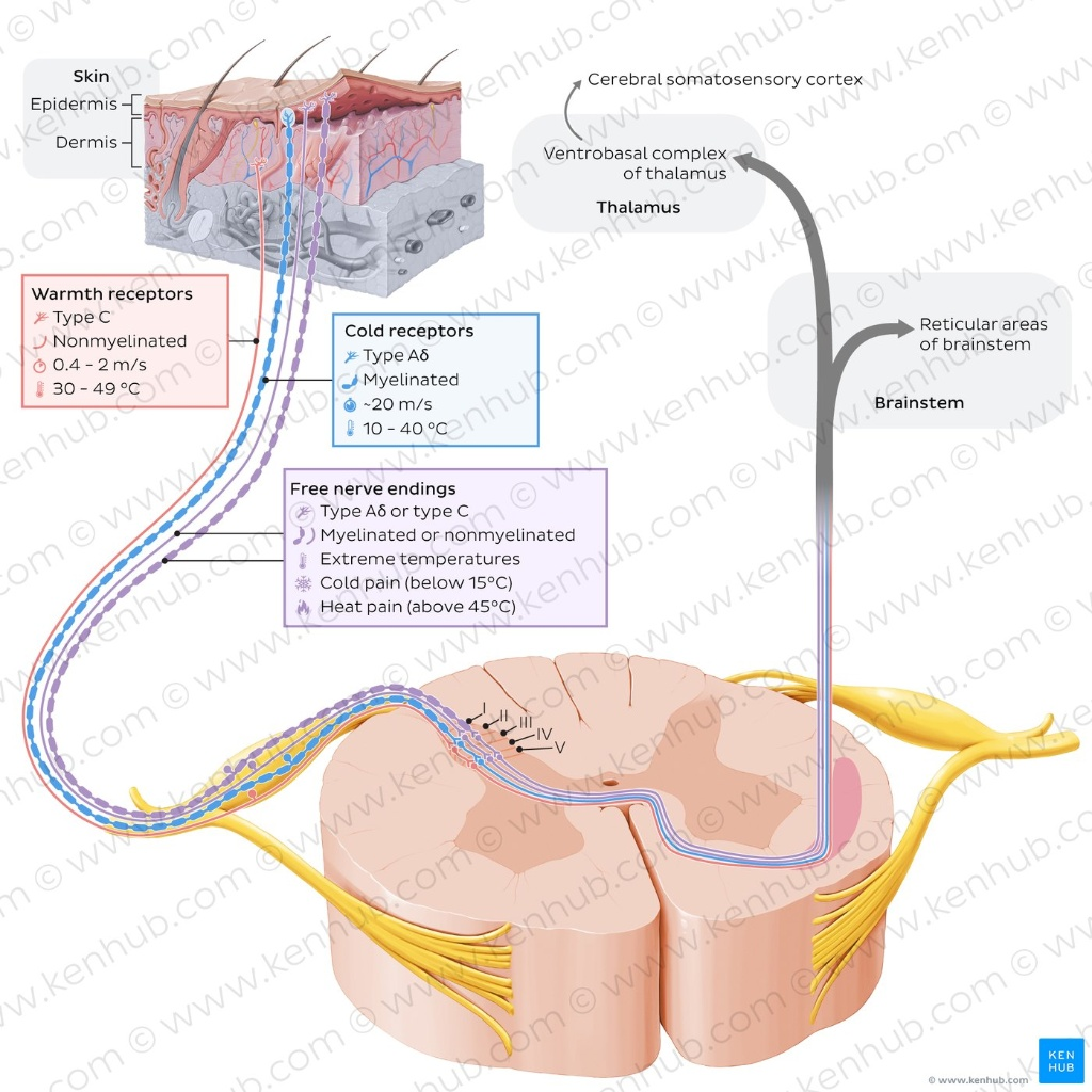
We have specialized sensors for hot and cold. The hot sensors range from body temperature (30°C) up to 45°C. They are fast-adapting, which is why a hot tub feels intensely hot at first but comfortable later. Anything above 45°C triggers the nociceptors (pain sensors), which are slow-acting and will continuously fire to warn you of tissue damage. If you've ever touched a motor controller running at 50°C, you know exactly what this feels like.
Cold receptors operate from body temp down to 10°C, helping us identify materials (metals wick heat faster than plastics) and detect wetness.
Robotic Equivalents for Sensor Networks
Researchers today evaluate several interesting tactile and temperature sensors, both individually and as arrays, to mimic the functionality of the human hand.
Forms
They primarily take four forms: thin-films, MEMS, molded materials, and textiles. Manufacturers create thin-film sensors by laminating many films together to form the sensing element and body. Micro-electro-mechanical systems (MEMS) are tiny integrated circuits that are fabricated using semiconductor techniques. They are lauded for their microscale and ability to be arranged into high-density arrays. Molded materials are custom polymers or gels with controlled material properties that enable the system to sense when the material moves or deforms. Finally, we have textiles with sensing technology woven into them. Let's dive into the sensing types to understand their strengths and weaknesses.
Sensing Elements
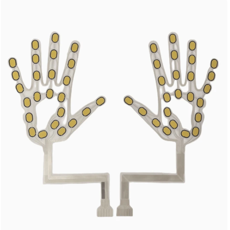
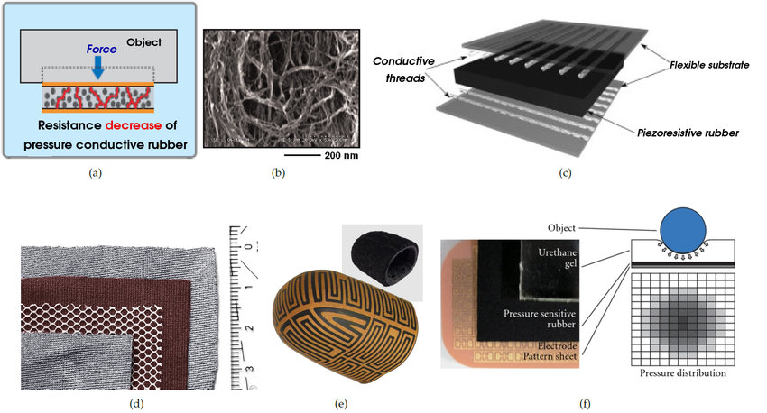
- Piezoresistive (FSR): Force-sensing resistors and piezoresistive tactile sensors measure changes in electrical resistance when pressure is applied. They are cost-effective, easy to integrate, and can be arranged into dense arrays or woven into textiles. Engineers have designed FSR arrays into the shape of a human hand for robotic grippers, and piezoresistive sensors in textile form enable flexible, conformable sensing surfaces. They excel at measuring static pressure and force distribution, though they may have limited bandwidth compared to piezoelectric sensors for high-frequency events.
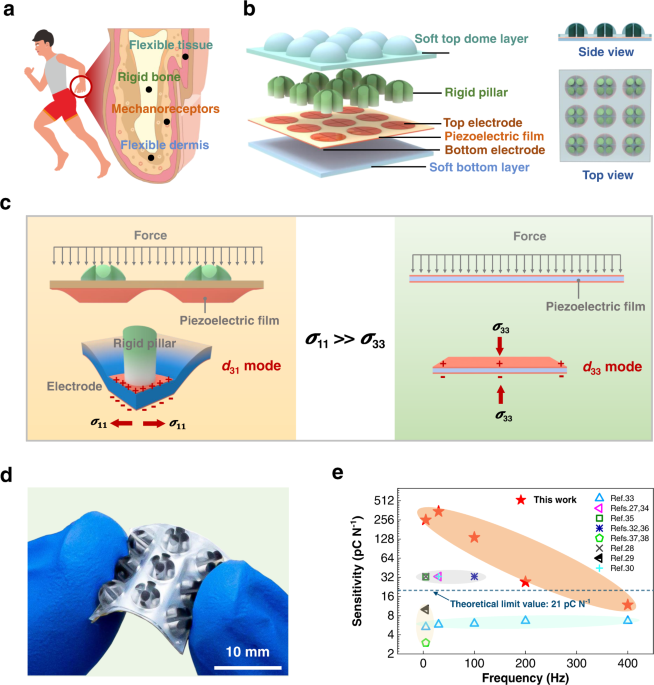
- Piezoelectric: These sensors generate a voltage spike when subjected to mechanical stress. Unlike static load measurements, piezoelectric sensors are insensitive to continuous pressure but highly sensitive to dynamic changes. This makes them excellent for detecting high-frequency events like slip or texture vibrations, rather than measuring the absolute force of a grasp. Piezoelectric sensing technology almost always uses MEMS construction, though engineers have successfully integrated it into thin-films as well.
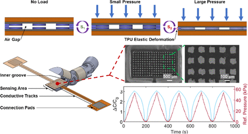
- Capacitive: Drawing from popular touchscreen technology, these sensors measure changes in capacitance as the dielectric layer between two conductive plates is compressed. They provide the dual ability to feel physical touch and sense the proximity of a nearby conductive object. They have moderate sensitivity and are almost always of thin-film construction, though arrays of capacitive MEMS devices embedded in molded materials are also prevalent.
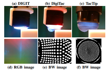
- Optical Touch Sensors: This technology combines a small camera with a molded material surface, typically a gel or polymer elastomer, that features internal tracking markers. The camera views the surface deformation from the inside to determine the applied force. It can capture high-grain detail for surface reconstruction and bulk deformation to calculate absolute pressure. Popular versions include GelSight and DIGIT. Constructed strictly as molded materials over optical hardware, the industry currently considers these the gold standard for texture and shape reconstruction.
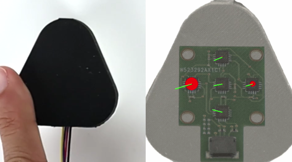
- Magnetic Sensors: These embed a passive magnet in a molded material and mount a Hall Effect sensor on a rigid or flex PCB. They are excellent for feature identification and tracking shear forces or edges, offering high sensitivity, low cost, and a rapid response time in the hundreds of Hertz. Systems like ReSkin or AnySkin feature replaceable magnetic outer skins, allowing for excellent serviceability. The downside is their susceptibility to electromagnetic interference from nearby motors or ferrous materials, meaning they cannot be the sole sensor type used in an unstructured environment.
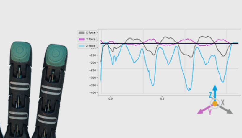
- Microbarometers: These sensors use localized changes in air or fluid pressure within a sealed cavity to measure tactile feedback. Manufacturers construct them by embedding MEMS barometers within a molded material. While most microbarometers are one-dimensional and only measure bulk pressure, advanced mechanical coupling designs, such as the SEED FTS system, enable some modern barometers to provide three-axis readings. The latest general-purpose robots from Sanctuary AI reportedly feature microbarometer tactile sensors.
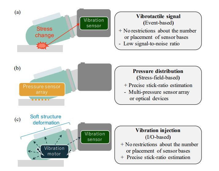
- Vibration Sensor: The traditional use of a vibration sensor is to passively detect slip vibrations and the resonant frequencies of the tools being manipulated. However, new approaches pair a MEMS vibration sensor with a vibration motor. The motor injects vibrations at specific frequencies into the fingertip, then the sensor reads the shifted return signal to determine skin deformation, applied force, and the acoustic impedance of the gripped object. These are exclusively MEMS constructions.
- Electrogel: Electrogels are molded materials that can change their resistivity, capacitance, and, potentially, inductance with deformation and temperature. Electrical impedance tomography (EIT) injects voltage at specific points on the skin and measures it at others. Algorithms then reconstruct these signals to map tactile pressure, damage, and temperature. Researchers propose them as the holy grail of electronic skins due to their monolithic molded construction and multimodal capabilities.
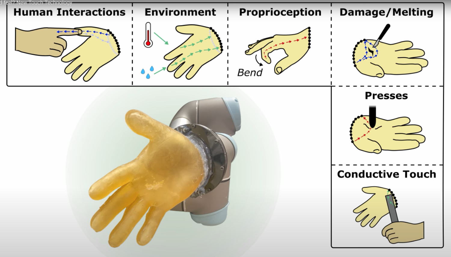
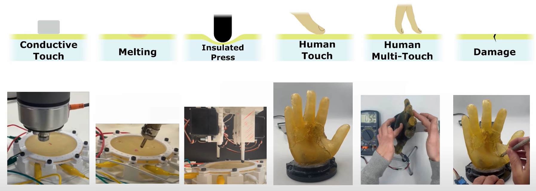
- Resistance Temperature Detectors (RTD): The RTD is king for localized temperature sensing. Primarily utilizing thin-film or MEMS construction, RTDs span the entire temperature range of human thermoreceptors and extend well into the nociceptor (pain) range, providing highly accurate and stable thermal data.
These technologies alone will likely not achieve the level of perception that human skin provides. Some sensors excel at measuring bulk pressure, others cover larger surface areas easily, and others are tuned for high-frequency vibrations. The human body effortlessly covers all of these modalities simultaneously. To achieve parity, we will likely integrate two or three construction types to cover 80 percent of what the human hand can do. These may be built into easily replaceable fingertips and palm pads, or they may eventually be covered by the Hail Mary approach of electrogels spanning the entire robotic hand.
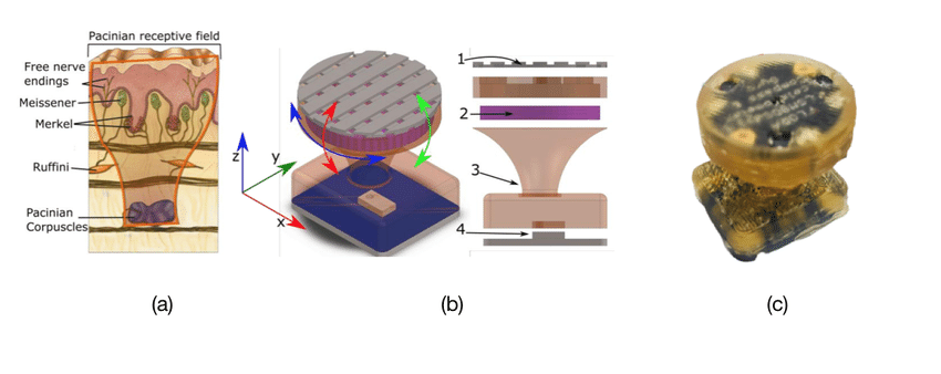
For high-frequency data capture, mimicking the Pacinian corpuscles in the 250 to 320 Hertz range, capacitive, resistive, or specialized MEMS devices, such as IMUs embedded in a molded skin array, are the best bets. This frequency-based spatial understanding is a massively unexplored area in robotics. It remains to be seen if advanced visual identification can overcome this gap. Can a robot recall enough object information visually to simply use touch for navigation rather than classification? The strong performance of purely vision-based object classifiers provides some basis for this. However, to truly bridge the gap, we will need massive, high-quality tactile training datasets to feed into VLA models, teaching robots the complex physical interactions that humans handle intuitively.
Conclusion
We like to focus heavily on the kinematics and muscles of robotic hands because they present a visible, obvious challenge. But we are missing a critical engineering hurdle: replicating the mechanical functions and dynamic tactile feedback of human skin.
One of the main reasons robots struggle with real-world manipulation today is this lack of physical awareness. Solving this problem without integrating tactile feedback will prove incredibly difficult. It's more than just registering pressure on a grid; it's about understanding contact states, material properties, and slip detection. To do real work in our unstructured world, robots need to understand how to grip, grasp, and manipulate objects even when they aren't looking directly at them.
Our mission at Morpheus Robotics is to make general-purpose robots useful in everyday life. We think tactile sense is a critical part of achieving viability in our world. We believe collecting large amounts of data with tactile sensing is one of the best ways to advance this mission. If you want to work on that same mission, come join us. Reach out via the contact page at morpheusrobotics.io.
Footnotes
[1] A biomechanical study detailing the ultimate tensile strength (UTS) and elastic properties of human skin under dynamic loads. Learn more. ↩
[2] A reference guide on Ultimate Tensile Strength, providing a baseline comparison for the mechanical limits of common engineering materials like rubber and ABS plastic. Learn more. ↩
[3] Research exploring the mechanical properties of the stratum corneum and how moisture plasticization transforms our skin into a highly compliant, high-traction grip surface. Learn more. ↩
[4] A clinical breakdown of mechanoreceptor densities in human glabrous (hairless) skin, specifically detailing the high concentration of Meissner corpuscles in our fingertips and palms. Learn more. ↩
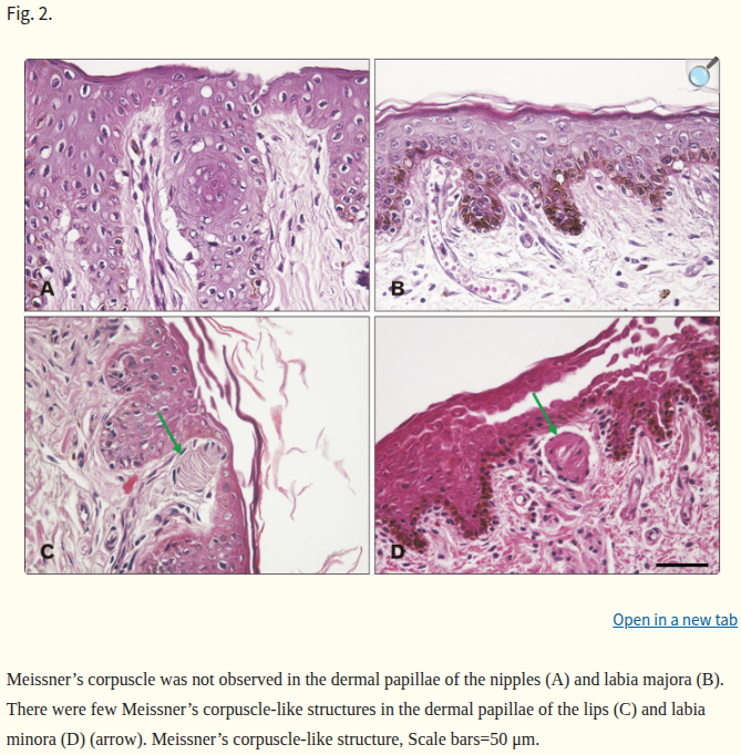
[5] A histological mapping of Pacinian corpuscles, highlighting their relative sparsity across the human hand compared to other mechanoreceptors. Learn more. ↩
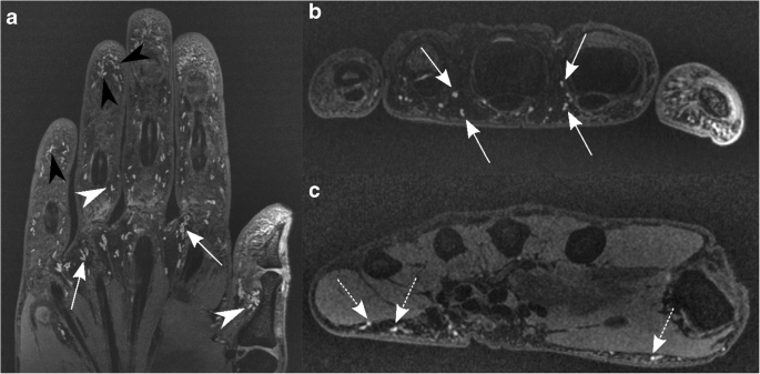
[6] A fascinating study on tool proprioception demonstrating how humans combine tactile vibration feedback with visual data to accurately estimate the length and spatial boundaries of a grasped object. For rigid objects, the performance drastically improved, but for non-rigid objects, the performance is no better than eyesight alone. Learn more. ↩